
| For Android: 4.1 and up | Guide: Positioning and Radiographic Anatomy of the Skull cheats tutorial |
| When updated: 2019-05-02 | Star Rating: 0 |
| Name: Positioning and Radiographic Anatomy of the Skull hack for android | Extension: Apk |
| Author: Medical Apps For Doctors | File Name: com.radiography.skullradiographicpositioning |
| Current Version: 1.0 | User Rating: Everyone |
| Downloads: 50- | Version: mod, apk, unlock |
| System: Android | Type: Education |


Watch Radiographic Positioning of the Skull video.

Watch RADT 210 Skull Positioning video.

Watch Basic Radiology of the Skull video.

Watch Radiographic Positioning (SKULL PART 1) video.

Watch 2016 WAERT Video Exhibit V2 Basics of skull positioning video.

Watch Anatomy of the Skull video.

Watch RADIOGRAPHIC POSITIONING (SKULL PART 2) video.

Watch Topography and Morphology of the Skull video.

Watch Anatomy and Positioning video.

Watch X-RAY OF ALL THE SKULL BONES , ANATOMY AND PHYSIOLOGY PART - 105 video.

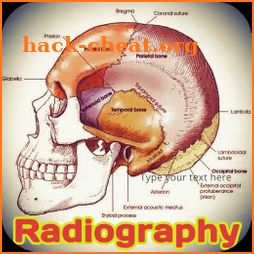
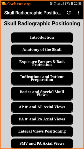
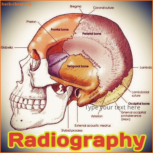
Skull Radiographic Positioning Positioning and radiographic anatomy of the skull Contents: Introduction Anatomy of the Skull Exposure Factors & Rad. Protection Indications and Patient Preparation Basics and Unique Skull Views AP 0° and AP Axial Views PA 0° and PA Axial Views Lateral Views Positioning SMV and PA Axial Views Sella Turcica Positioning Facial Bones Positioning I Facial Bones Positioning II Facial Bones Positioning III Facial Bones Positioning IV - Skull Radiographic Positioning Facial Bones Positioning V Nasal Bones positioning Orbit Positioning I - Skull Radiographic Positioning Orbit Positioning II Paranasal Sinuses Positioning I Paranasal Sinuses Positioning II Mandible Positionig I Mandible Positioning II Mandible Positioning III TMJ Positioninig I TMJ Positioninig II Mastoids Process Positioning I Mastoids Process Positioning II - Skull Radiographic Positioning Positioning and radiographic anatomy of the skull 1. Positioning and Radiographic Anatomy of the Skull 2. RADIOGRAPHIC ANATOMY Skull As with another body parts, radiography of the skull requires a awesome understanding of all similar anatomy. The anatomy of the skull is very complex, and specific attention to detail is needed of the technologist. The skull, or bony skeleton of the head, rests on the superior end of the vertebral column and is divided into two main sets of bones—the 8 cranial bones and the 14 facial bones. 3. CRANIAL BONES (8) The eight bones of the cranium are divided into the calvaria (skullcap) and the floor. Each of these two places primarily consists of four bones: Calvaria (Skullcap) 1. Frontal 2. Right parietal 3. Left parietal 4. Occipital Floor 5. Right temporal 6. Left temporal 7. Sphenoid (sfe′-noid) 8. Ethmoid (eth′-moid) 4. POSITIONING CONSIDERATIONS Erect against Recumbent Projections of the skull may be taken with the patient in the recumbent or erect position, depending on the patient's condition. Photos can be obtained in the erect position with the use of a standard x-ray table in the vertical position or an upright Bucky. The erect position allows the patient to be quickly and easily positioned and permits the use of a horizontal beam. A horizontal beam is important to visualize any existing air-fluid levels within the cranial or sinus cavities 5. Patient Comfort Patient motion almost always results in an unsatisfactory photo. During skull radiography, the patient's head must be placed in precise positions and held motionless long enough for an exposure to be obtained. Always remember that a patient is attached to the skull that is being manipulated. Every effort could be created to create the patient's body as comfortable as possible, and positioning aids such as sponges, sandbags, and pillows could be used if required. Except in cases of severe trauma, respiration could be suspended during the exposure to assist prevent blurring of the photo caused by breathing movements of the thorax. This is especially necessary when the patient is in a prone position. 6. Hygiene Cranial and facial radiography may require the patient's face to be in direct contact with the technologist's hands and the table/upright Bucky surface. Therefore, it is necessary that proper handwashing techniques and surface disinfectants be used before and after the examination. 7. Exposure Factors The principal exposure factors for radiography of the skull contain the following: •Medium kV (65 to 85 kV film-based) (70 to 80 kV digital radiography [DR] and computed radiography [CR]) •Little focal spot <250 mA (if equipment allows) •Short exposure time. Find out more in the apk. Skull Radiographic Positioning If you like the apk please leave a positive review and share with your colleagues. Thank you for using this app. Skull Radiographic Positioning


 War Master : Strategy Battle
 playpod
 Kela Pro
 Potty with Pull-Ups ft. Disney
 Learn AI & Chat GPT: Gen AI X
 The Soundscape: Piano Run
 Cocobi Ice Cream Truck - Kids
 The Visitor
 RedWheel
 Bubble Cash
 BFF14-Halloween Pumpkin Castle Hacks
 AI Story Generator AI Writer Hacks
 Stickman Empire : Strategy War Hacks
 Tekmetric Mobile Hacks
 Grab Throw : Hit Annoying Men Hacks
 Top Dogs Hacks
 Embody Hacks
 Treasure Chest Clicker - Idle Hacks
 PegIdle Hacks
 PDF Reader - Document Viewer Hacks
Share you own hack tricks, advices and fixes. Write review for each tested game or app. Great mobility, fast server and no viruses. Each user like you can easily improve this page and make it more friendly for other visitors. Leave small help for rest of app' users. Go ahead and simply share funny tricks, rate stuff or just describe the way to get the advantage. Thanks!
Welcome on the best website for android users. If you love mobile apps and games, this is the best place for you. Discover cheat codes, hacks, tricks and tips for applications.
The largest android library
We share only legal and safe hints and tricks. There is no surveys, no payments and no download. Forget about scam, annoying offers or lockers. All is free & clean!
No hack tools or cheat engines
Reviews and Recent Comments:

Tags:
Positioning and Radiographic Anatomy of the Skull cheats onlineHack Positioning and Radiographic Anatomy of the Skull
Cheat Positioning and Radiographic Anatomy of the Skull
Positioning and Radiographic Anatomy of the Skull Hack download